Plant Cells vs Animal Cells Venn Diagram: A KS3 Biology Guide
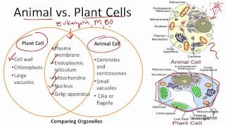
Understanding the similarities and differences between plant cells and animal cells is a core part of KS3 biology. One of the most effective visual tools for this comparison is a Venn diagram. By placing the unique features of each cell type on the outer circles and the shared organelles in the overlapping section, students can quickly grasp the essential concepts required for exams and classroom discussions.
Why Use a Venn Diagram?
A Venn diagram helps learners:
- Organise information in a clear, side‑by‑side format.
- Identify common organelles such as the nucleus, mitochondria, and ribosomes.
- Highlight unique structures like the cell wall in plant cells or centrioles in animal cells.
- Prepare for questions that often appear in KS3 assessments and online quizzes, for example on Braingenie.
Key Differences Between Plant and Animal Cells
When constructing the outer sections of the Venn diagram, list the organelles and features that are exclusive to each cell type.
- Cell Wall – A rigid layer of cellulose that provides structural support to plant cells. Animal cells lack a cell wall.
- Chloroplasts – Contain chlorophyll for photosynthesis; absent in animal cells.
- Large Central Vacuole – Stores water, nutrients, and waste; helps maintain turgor pressure in plants.
- Centrioles – Present in animal cells and play a role in cell division; typically missing in most plant cells.
- Lysosomes – More common in animal cells for digestive processes, though some plant cells have similar structures.
Common Features (Overlap in the Venn Diagram)
The intersecting area of the diagram should contain organelles that both plant and animal cells share:
- Nucleus – Holds genetic material (DNA) and controls cell activities.
- Plasma Membrane – Regulates the movement of substances in and out of the cell.
- Mitochondria – The “powerhouses” that generate ATP through cellular respiration.
- Endoplasmic Reticulum (ER) – Rough ER (with ribosomes) for protein synthesis; smooth ER for lipid synthesis.
- Golgi Apparatus – Modifies, sorts, and packages proteins and lipids.
- Ribosomes – Sites of protein synthesis, either free‑floating or attached to the ER.
![Plant and Animal Cells [KS3 SCIENCE]](https://i.ytimg.com/vi/FZtMCxFJYbw/mqdefault.jpg)